암 진단에서 조직 생검은 오랫동안 ‘확실한 진단 수단’으로 여겨져 왔다. 그러나 실제로는 조직 절개나 굵은 바늘 삽입이 수반되어 환자에게 상당한 통증과 흉터, 출혈, 감염 위험을 안긴다. 특히 뇌, 폐, 췌장과 같은 민감한 장기에서는 반복적인 검사 자체가 어렵고 심지어 위험하기까지 하다. 진단 결과를 기다리느라 치료 개시가 지연될 수 있고, 이는 결국 예후에도 직접적으로 부정적인 영향을 미친다. 이처럼 전통 생검은 진단의 안정성에는 강점을 보이나, 그 효율성과 환자 편의성에는 큰 한계를 지니고 있으며, 의료 현장에서는 오랫동안 조직 손상 없이 빠르고 안전하게 검체를 확보할 수 있는 기술에 대한 갈망이 지속되어 왔다.
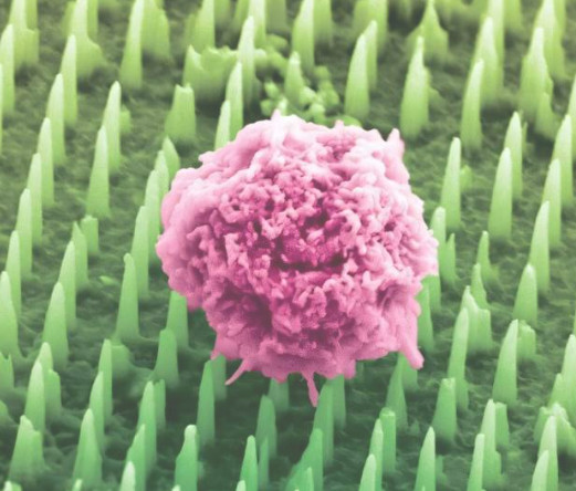
초미세 실리콘 바늘이 만든 패치
킹스 칼리지 런던(King’s College London) 연구팀은 이러한 필요에 응답하여 무려 수백만 개의 수십~수백 나노미터 실리콘 나노‑니들을 플라스터 형태의 얇은 패치에 집적하였다. 머리카락보다 약 1,000배 가는 이 나노‑니들은 조직 표면에 가볍게 붙이는 것만으로도 세포 외막을 부드럽게 관통하여 지질, 단백질, mRNA 등 다양한 생체 분자들을 포집한다. 절개나 깊은 삽입을 하지 않으므로 통증과 출혈은 거의 없으며, 생체적합성 또한 높아 부작용 우려가 낮다. 이 패치를 통해 의료진은 조직을 수동적으로 손상하지 않고도 고정밀 분자 샘플을 획득할 수 있으며, 이는 암 진단의 본질을 재정의하는 혁신적 전환점이라 할 수 있다.
인공지능(AI) 기반 실시간 진단
포집된 분자 데이터는 고성능 질량분석 장비와 인공지능 기반 알고리즘으로 정교하게 분석되어, 20분 이내에 암의 존재뿐만 아니라 종양의 유형과 조직 등급까지 평가할 수 있다. 전통 생검이 수일 걸리던 과정과 비교하면 시간 효율은 말할 것도 없으며, 20분 내 실시간 결과는 수술 중에도 즉시 활용 가능하다. 예를 들어 뇌수술 중 의심 병변에 패치를 부착한 뒤 분석 결과를 반영하여 절제 범위나 수술 방향을 즉각 조정할 수 있으며, 이는 수술 성공률을 높이고 불필요한 조직 손상을 줄이는 효과를 가져올 것이다.
반복 가능한 분자 지도
나노‑니들 패치는 조직을 건드리지 않기 때문에 동일 부위에 여러 차례 부착한 후 분자 샘플을 지속적으로 수집할 수 있다. 이로 인해 치료 전후, 회복기, 재발 직전 등 다양한 시점에서 분자 상태의 변화를 시각적으로 기록할 수 있으며, 이를 통해 분자 지도(molecular map)라고 불리는 시공간적 분석이 가능하다.
여기서 분자 지도란 환자의 특정 조직이나 종양이 시간에 따라 분자적 특성이 어떻게 변화하는지를 시각화한 분석 자료를 말한다. 예를 들어 치료 효과가 어떻게 나타나는지, 약물 반응은 어떠한지, 재발 가능성은 어느 정도인지 등을 시간 순서대로 보여주는 정보로, 매우 정밀한 맞춤형 치료 설계에 중요한 자료가 된다. 이 지도는 종양의 성장, 약물 반응, 재발 가능성과 같은 중요한 생체 변화를 시간 축 위에 관찰하게 해 줄 것이며, 의료진은 이를 토대로 더 정밀하고 개인화된 치료 전략을 수립할 수 있다.
전임상 단계에서 얻은 성과
전임상 연구에서는 인간 및 동물의 뇌종양 조직 23건을 대상으로 나노‑니들 패치를 적용하였다. 그 결과 교모세포종과 수막종 등의 다양한 종양 유형을 정확하게 구분했으며, 분석 결과는 20분 이내에 도출되었다. 이 성과는 단순한 진단 도구를 넘어 ‘동일 조직의 시공간적 분자 정보를 동시에 분석하고 지도화할 수 있다’는 공간생물학(spatial biology) 관점의 가능성을 입증했다. 수술실에서 즉시 적용 가능한 분자 기반 진단 도구의 첫 발걸음이 마련된 셈이다.
확장성 높은 제조 기술
이 기술은 반도체 리소그래피 공정 기반으로 제작되어 컴퓨터 칩과 동일한 방식으로 대량 생산이 가능하다. 이로 인해 제조 단가를 낮추고, 의료 현장 도입 장벽을 낮출 수 있다. 또한 이 패치는 밴디지, 내시경 팁, 콘택트렌즈 등 다양한 의료기기에 쉽게 통합될 수 있어 구강, 호흡기, 피부, 뇌 등 여러 장기로의 확장이 용이하다. 더 나아가 알츠하이머나 자가면역질환 진단 등에도 적용 가능성이 있어, 응용 범위는 매우 광범위하다.
임상 적용을 위한 과제들
현재 기술은 전임상 단계에 머물러 있다. 향후에는 인체 대상 임상시험(1·2상)에서 안전성과 유효성을 입증해야 한다. 동시에 인공지능 알고리즘 인증, 의료기기 허가, 보험 수가 책정, 진단 지침 반영 등 제도적 기반 구축도 필수이다. 또한 각 질환의 특성에 맞는 맞춤 설계와 생산 공정 고도화, 의료진의 현장 활용 역량 확보를 위한 교육 시스템 구축도 동반되어야 한다.
암 진단의 패러다임이 바뀔 것이다
결론적으로, 나노‑니들 패치는 비침습·무통·실시간·반복 가능이라는 특징으로 암 진단의 방식 자체를 바꿀 것이다. 환자는 진단의 고통과 불안을 덜고 효율적인 검사를 받을 수 있으며, 의료진은 20분 내 분자 기반 실시간 진단을 통해 보다 정확한 치료 전략을 설계할 수 있다. 이는 특히 수술실에서 효과적일 것이며, 결과적으로 환자의 예후와 삶의 질을 크게 향상시킬 것이다. 향후 5년 내 임상 적용이 실현된다면, 암뿐 아니라 난치성 질환 전반의 진단-치료 체계가 정밀하고 개인화된 의료 형태로 전환되며 의료 패러다임 전체가 혁신될 것이다.
* Reference
Nature Nanotechnology – June 16, 2025, "Nanoneedle patch offers painless alternative to traditional cancer biopsies," King’s College London.
Innovation in Non-Invasive Diagnostics: The Emergence of the Nanoneedle Patch
Beyond the Limitations of Traditional Biopsy
In cancer diagnostics, tissue biopsy has long been considered the “gold standard” for definitive diagnosis. However, in practice, it involves tissue excision or the insertion of large needles, causing patients significant pain, scarring, bleeding, and risk of infection. In particularly sensitive organs such as the brain, lungs, and pancreas, repeated biopsies are difficult and sometimes even dangerous. Diagnostic delays while waiting for biopsy results can negatively affect prognosis. Thus, while traditional biopsies offer diagnostic accuracy, they pose significant limitations in efficiency and patient comfort. For years, the medical community has sought technologies that can obtain samples quickly and safely, without tissue damage.
A Patch Made of Ultra-Fine Silicon Needles
A research team at King’s College London has responded to this need by embedding millions of ultra-thin silicon nanoneedles (tens to hundreds of nanometers in diameter) into a thin, plaster-like patch. These nanoneedles, roughly 1,000 times thinner than a human hair, gently penetrate the extracellular membrane simply by being placed on the tissue surface, collecting various biomolecules such as lipids, proteins, and mRNA. Since no incision or deep insertion is required, there is little pain or bleeding, and the patch demonstrates high biocompatibility with minimal side effects. This enables clinicians to obtain high-precision molecular samples without damaging the tissue, representing a transformative advancement in cancer diagnostics.
AI-Powered Real-Time Diagnosis
The collected molecular data is precisely analyzed using high-performance mass spectrometry and AI-based algorithms, enabling diagnosis within 20 minutes. Not only can the presence of cancer be detected, but tumor type and tissue grading can also be assessed. Compared to traditional biopsy processes that take several days, this rapid turnaround allows for real-time application even during surgery. For instance, during brain surgery, the patch can be applied to suspicious lesions, and the results used to immediately adjust the surgical scope and direction, increasing success rates while minimizing unnecessary tissue damage.
Repeatable Molecular Mapping
Because the nanoneedle patch does not disrupt the tissue, it can be applied repeatedly to the same area, allowing for continuous collection of molecular samples. This enables visualization of molecular changes over time—before and after treatment, during recovery, or prior to recurrence—forming what is known as a “molecular map.” Such a map allows clinicians to observe important biological changes such as tumor growth, drug response, and recurrence potential over time, and to develop more precise, personalized treatment strategies based on these insights.
Preclinical Achievements
In preclinical studies, nanoneedle patches were applied to 23 human and animal brain tumor samples. The patches accurately distinguished various tumor types, such as glioblastoma and meningioma, with results produced within 20 minutes. This achievement goes beyond the role of a simple diagnostic tool, demonstrating the potential of spatial biology: the ability to simultaneously analyze and map the spatial and temporal molecular information of the same tissue. It marks a critical first step toward molecular-based diagnostic tools that can be used immediately in operating rooms.
Scalable Manufacturing Technology
This technology is manufactured using semiconductor lithography processes, allowing for mass production using the same methods as computer chips. This lowers manufacturing costs and reduces barriers to clinical adoption. Additionally, the patch can be easily integrated into various medical devices such as bandages, endoscopic tips, and contact lenses, enabling application to the oral cavity, respiratory system, skin, brain, and other organs. Moreover, the patch holds promise for diagnosing conditions like Alzheimer’s disease and autoimmune disorders, greatly broadening its potential applications.
Challenges for Clinical Implementation
Currently, the technology remains in the preclinical stage. Moving forward, it must demonstrate safety and efficacy in human clinical trials (Phases 1 and 2). Institutional groundwork—including AI algorithm validation, medical device approval, insurance reimbursement codes, and incorporation into diagnostic guidelines—is also essential. Furthermore, it will require customized designs tailored to each disease, refinement of production processes, and the establishment of training systems to ensure healthcare professionals can use the technology effectively in clinical settings.
A Paradigm Shift in Cancer Diagnostics
In conclusion, the nanoneedle patch—characterized by non-invasiveness, painlessness, real-time analysis, and repeatability—is poised to revolutionize the very approach to cancer diagnostics. Patients will experience less pain and anxiety while undergoing efficient testing, and clinicians will be able to design more accurate treatment strategies through molecular-based real-time diagnostics in just 20 minutes. This will be particularly valuable in surgical settings, ultimately improving patient outcomes and quality of life. If clinical implementation is realized within the next five years, it could drive a broad transformation of diagnostic and therapeutic systems—not only for cancer but also for intractable diseases—toward precision and personalized medicine, ushering in a new era in healthcare.
* Reference
Nature Nanotechnology – June 16, 2025, "Nanoneedle patch offers painless alternative to traditional cancer biopsies," King’s College London.